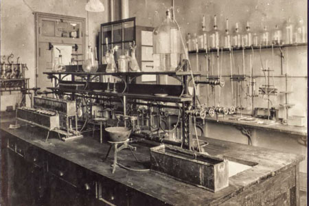
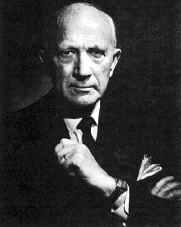
David Hunter Hubel, FRS, Nobel Laureate, neurobiologist (born 27 February 1926 in Windsor, ON; died 22 September 2013 in Lincoln, Massachusetts). Dr. David Hubel advanced our understanding of how our eyes transmit and reconstitute the images we see moment by moment to our brains. He was a research scientist who used innovative craftsmanship to design and create the instruments and procedures he used to conduct studies on the visual cortex of the brain. His meticulous observations revolutionized cognitive neuroscience and his pioneering approach to the recording of individual neuronal cells propelled the field forward.

Early Life
David Hubel’s parents, Jesse Hervey Hubel and Elsie Mabel Hunter, moved from Detroit, Michigan to Windsor, Ontario for his father’s employment as a chemical engineer at the Windsor Salt Company. David Hubel, who was born in Windsor in 1926, was Canadian by birth and a US citizen through his parents. (See also Canadian Citizenship.)
In 1929, Hubel’s family moved to Montreal, Quebec. Hubel was interested in science from a young age and his hobbies included chemistry and electronics. His mother was fascinated with electricity and his father, a chemical engineer gifted Hubel a chemistry set. David Hubel was a student at Strathcona Academy in Montreal’s Outremont borough for 12 years. He often recalled his outstanding teachers before entering McGill University in Montreal.
Education and Early Career
After obtaining an honours undergraduate degree in mathematics and physics at McGill University in 1947, David Hubel was accepted as a medical student at McGill. His passion for brain research was kindled at this time (see Neuroscience). In his second year, he met Wilder Penfield, the founder of the Montreal Neurological Institute (MNI), who suggested that Hubel meet with the MNI’s Herbert Jasper. Over the summer, Hubel worked in Jasper’s physiology lab doing electronics. Hubel earned his MDCM in 1951 and pursued his neurology residency and internship at the Montreal General Hospital. Hubel continued as a research fellow with Jasper, learning how to perform and interpret clinical EEG (electroencephalography), which Jasper used to study electrical activity of the brain. During this time, Jasper assigned Hubel the topic of the visual system for his neurophysiology seminar series. Hubel uncovered Steven Kuffler’s 1952 paper on neurons in the retina that launched his life-long interest.
Teaching EEG methodology to a visiting neurologist from Johns Hopkins propelled Hubel to accept an offer from Hopkins for a neurology residency in 1954. After his first year, and to avoid the doctors’ draft he volunteered for the United States Army to be assigned to the Walter Reed Army Institute of Research in Washington DC. (See also Korean War.) By 1957, he made an innovative medical breakthrough at the Walter Reed. Hubel crafted a tungsten wire micro-electrode by sharpening it using an alternating current in a potassium nitrite bath. Finally, insulating the micro-electrode with a concentrated lacquer called “Insulex” enabled successful single-cell recordings in the brain.
Identifying his former student’s ingenuity, Herbert Jasper came to Walter Reed to learn how to make such micro-electrodes. Jasper then used the technology for his own experiments, enabling him to make single-cell recordings in the brain. Hubel recognized the need for a micro-electrode advancer to position his tungsten micro-electrode for single cell recordings. He took a night school course in machining to make the hydraulic micro-electrode advancer. His tungsten electrodes and micro-electrode advancer were critical for his subsequent discoveries.
Key Discoveries
At Walter Reed in 1957, David Hubel began to study the visual cortex of the brain through his recording of single cells. After turning the lights off and on, he noticed several different cells had responded to light stimuli, confirming the work of others. With his micro-electrode, he discovered that a previously unresponsive cell reacted preferentially to visual stimuli of motion (hand waving).
Another scientist who came to Walter Reed to learn how to make Hubel’s tungsten micro-electrodes was Torsten Wiesel from Stephen Kufflers’s laboratory at the Johns Hopkins Wilmer Eye Institute. At the invitation of Vernon Mountcastle, Hubel moved back to Johns Hopkins in 1958 to pursue a post-doctoral fellowship. However, a serendipitous turn of events directed Hubel’s career towards Stephen Kuffler, often regarded as the “father of modern neuroscience”. In Kuffler’s laboratory, Hubel began his formal collaboration with Torsten Wiesel.
In 1959, Hubel and Wiesel found three neighbouring responsive cells with parallel receptive fields which responded in such a way as to imply that the visual cortex as a whole was organized in columns. They discovered cells specialized for responding to straight lines and different orientations of these lines.
With Stephen Kuffler’s laboratory, Hubel with Wiesel moved to Harvard University in 1959 where they later became founding members of the Department of Neurobiology. At Harvard, Hubel and Wiesel continued their collaborative research.
In their 1962 landmark paper "Receptive fields, binocular interaction and functional architecture in the cat’s visual cortex”, they deduced a columnar organization of these cells known as orientation columns. They further discovered that the orientation columns were organized with those responsive to the right eye adjacent to those from the left eye. These were referred to as the ocular dominance columns. The signals were combined in binocular cells to give stereoscopic depth. By using electrodes to make anatomically precise lesions, they were able to map the locations of the responsive cells they had discovered in the visual cortex.
Their later research on new-born kittens revealed that sensory-deprivation of one eye led to a marked decline of its associated ocular dominance column neurons in the primary visual cortex that is irreversible even after eye opening. The visual cortex neurons of the closed eye were not lost but had rather shifted their responses to the ocular dominance column for the open eye. This influenced physicians to ensure early treatment after birth for similar conditions, such as the early treatment of cataracts in the eyes of newborns.
For these discoveries, David Hubel and Torsten Wiesel’s common lifelong interest in the visual system culminated in the Nobel Prize in Physiology or Medicine in 1981. (See also Nobel Prizes and Canada.)
Career Highlights and Legacy
David Hubel’s success has been attributed to his mechanical inventiveness and perseverance. Hubel’s innovation of the tungsten micro-electrode enabled single-cell recordings of neurons. This was a major advance over the use of glass pipettes. The innovation of the hydraulic electrode micro-drive included a way to seal the electrode advancer to the cortex for stable recording.
Hubel remained at Harvard for the rest of his career. After officially retiring, he continued to teach and encouraged students be hands-on.
Did you know?
According to Eric Kandel, another winner of the Nobel Prize in Physiology or Medicine, David Hubel and Torsten Wiesel’s body of work “stands as one of the great biological achievements of the 20th century.” – The New York Times, 2013
With his wife Ruth, Hubel established undergraduate science research scholarships at Dalhousie University, Wellesley College, and the University of Windsor. David Hubel is interred at the Mount Royal Cemetery in Montreal.
Personal Life
David Hubel was a lover of music, an interest that began with piano lessons before he could read. His passion for music extended to his joining a McGill University choir where he met his future wife, Ruth Izzard. In his biography for the Nobel Prize, Hubel indicated that his interest in music included the piano, recorders and the flute. His biography also noted that he enjoyed woodworking, astronomy, skiing, tennis, squash, and learning different languages.
Honours and Awards
As well as winning numerous prestigious awards, David Hubel was also the recipient of 13 honorary degrees.
- Fellow, American Academy of Arts and Sciences (1965)
- Fellow, National Academy of Sciences (1971)
- Inaugural Rosenstiel Award, Brandeis University (1971)
- Karl Spencer Lashley Award, American Philosophical Society (1977)
- Louisa Gross Horwitz Prize, Columbia University (1978)
- Dickson Prize in Medicine, University of Pittsburgh (1980)
- Nobel Prize in Physiology and Medicine, Nobel Assembly at Karolinska Institute (1981)
- Foreign Member, Royal Society of Great Britain (1982)
- Fellow, American Philosophical Society (1982)
- Ralph W. Gerard Prize in Neuroscience, Society for Neuroscience (1993)
- Inductee, Canadian Medical Hall of Fame (2006)
Key Terms: David H. Hubel
Visual Cortex – The posterior region of the brain which receives, integrates, and processes visual information from our retinas. The visual information is integrated and sent to other regions of the brain for rapid recognition of objects and patterns with no conscious effort.
Visual Receptive Field – The region of the eye which, when stimulated, evokes a sensory response in a neuron.
Cataracts in Children – Cataracts in both eyes may be responsible for between 5 per cent to 20 per cent of childhood blindness globally. Due to the basic research of David Hubel and Torsten Wiesel, surgeons now remove cataracts from newborns and young children with resultant good visual outcomes that would otherwise lead to blindness.
Micro-electrode – A very small electrode that can be inserted into living tissue. Micro-electrodes can be used to record the electrical activity of the brain or a single cell.
EEG (electroencephalography) – Method used to record electrical activity of the brain using electrodes attached to the scalp.

 Share on Facebook
 Share on X
 Share by Email
 Share on Google Classroom


